This page contains automated translations.

The Academic Expert Program Cytology offers biomedical scientists highly specialized continuing education in the field of cytology. You will deepen your expertise in gynecological and extragenital cytology, learn about modern molecular pathological methods, and acquire in-depth knowledge of digital pathology— a field with a promising future. Lay the foundation for your specialized career while simultaneously strengthening your diagnostic confidence.

Department
Health Sciences

Highlights

  • in-depth study of gynecological cytology and extragenital cytology

  • molecular pathology

  • digitalization, quality management

  • case management

  • seamless progression to the Master’s Program (CE) Health Studies is possible


    Facts

    Final degree

    Academic Expert in Cytology

    Duration of course
    2 semesters
    Organisational form
    part-time

    Tuition fee

    Once payment € 7.3501

    + ÖH contribution + optional costs for additional services, if applicable2

    ECTS
    60 ECTS
    Language of instruction
    German

    Currently no application possible

    Study places

    20

    1 You can find an alternative model for semester payments under ‘Costs’.
    2 The costs depend on the additional services selected from the University of Applied Sciences Campus Vienna, such as work clothing, licenses, tutorials, or excursions.
    Insights into the Program

    <
    >
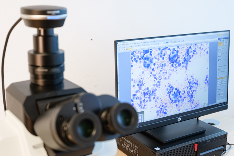
    Im Vordergrund ein Mikroskop; im Hintergrund ein Monitor.

    Digital microscopy: Microscopic specimens are scanned at high resolution and made available in digital form. This allows the specimens to be viewed, analyzed, and shared with others via a computer, regardless of their location.

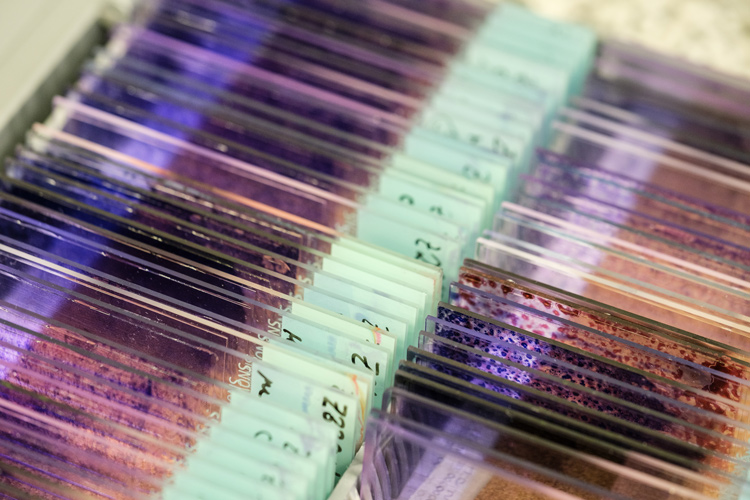
    Zahlreiche Objektträger für den Gebrauch unter einem Mikroskop.

    Archival storage box: Storage of microscope slides with stained smear slides to ensure their long-term availability for documentation and quality assurance purposes.

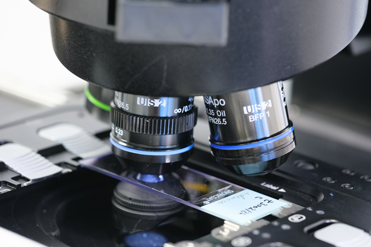
    Nahausnahme eines Objektträgers in einem Mikroskop

    Microscopic evaluation: The cytology specimen is screened using a meandering motion to ensure complete coverage of the entire specimen and reliable detection of abnormal cells.

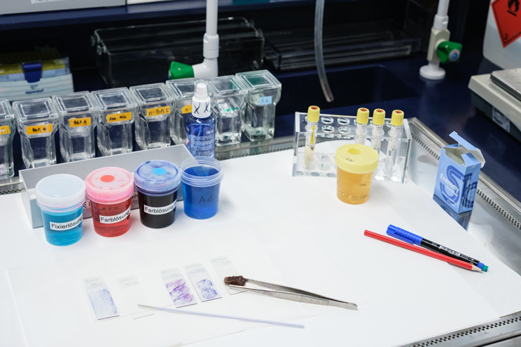
    Laborumgebung

    May-Grünwald-Giemsa Rapid Staining: This staining method makes cells and other components of urine obtained via a catheter visible, allowing for their microscopic evaluation.

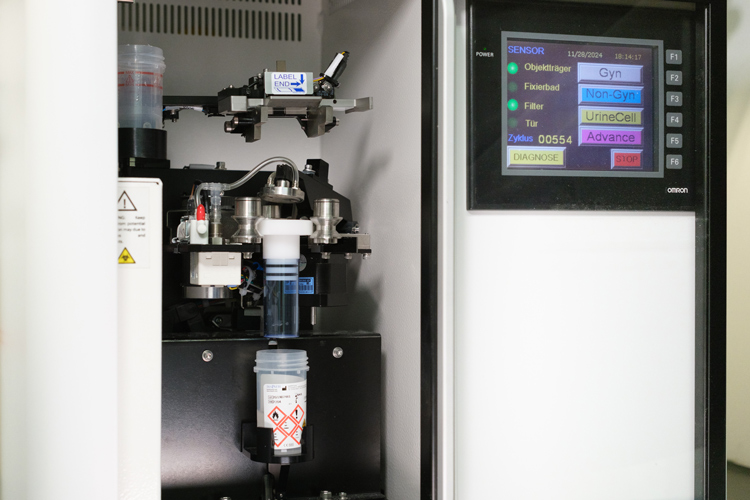
    Thin-layer cytology: Preparation of a genital cell specimen using the thin-layer technique. In this process, the cells are evenly distributed as a monolayer on a microscope slide to facilitate accurate microscopic evaluation.

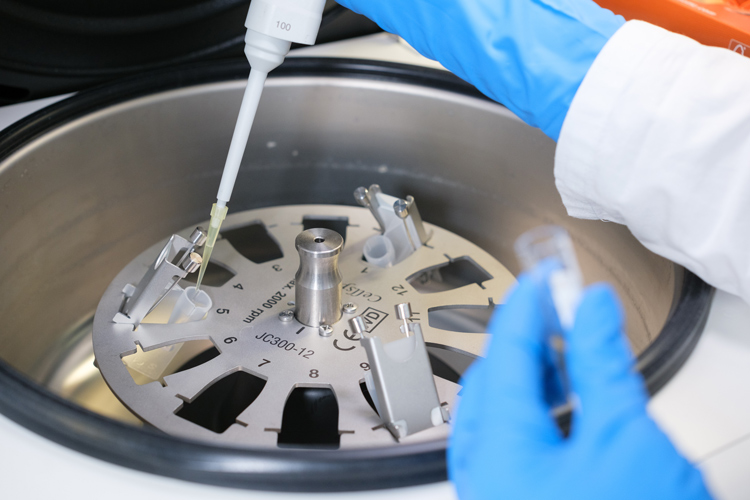
    Preparation of a cytospin: Cells from samples with low cell counts are concentrated on a small area of a microscope slide using a cytocentrifuge.


    Before the studies

    Are you already working as a biomedical scientist and would like to gain further qualifications in cytology? At a time when digitalization is bringing about lasting changes in diagnostics, you’ll prepare yourself for the implementation of artificial intelligence in everyday laboratory work. This program is ideal for you if you want to expand your expertise in the field of precision diagnostics while simultaneously building skills in quality assurance and research. Whether in the clinical setting or as a freelancer—here you’ll deepen your expertise.

    Why you should study with us

    Unique specialization

    One of the few programs in continuing education in the DACH region that combines classical cytology with the requirements of digital transformation and AI.

    Experts with practical experience

    Learn from leading professionals in the field who incorporate latest case studies and real-world laboratory scenarios into their teaching.

    Flexibility for working professionals

    Thanks to the combination of online lectures, interactive self-study modules, and concentrated on-campus sessions, the academic expert program can be seamlessly integrated into your daily work routine.

    Relevant admission requirements

    The relevant admission requirements for this academic expert program are:

    • a completed Bachelor’s degree in biomedical science in accordance with FH-MTD-AV or
    • a diploma in biomedical science in accordance with FH-MTD-AV or
    • an equivalent or higher-level final degree from a domestic or foreign post-secondary or tertiary educational institution in the field of biomedical science.

    If a post-secondary degree (e.g., diploma) is presented, the following must also be provided:

    • a general university entrance qualification, evidenced by an
      • Austrian secondary school leaving certificate (AHS, BHS, higher education entrance qualification) or
      • a certificate of a limited higher education entrance qualification for the respective field of study or
      • an equivalent foreign certificate
    • or, alternatively, by subject-specific supplementary exams in German (B2).

    Additional requirements

    In addition, proof of valid registration in the Health Professions Register must be provided.

    Language requirements for admission

    The required language level according to the Common European Framework of Reference for Languages (CEFR) is at least

    • German - level B2.

    Legalization of foreign documents

    Applicants may require legalization of documents from countries other than Austria in order for them to have the evidential value of domestic public documents. Information on the required legalizations can be found here in PDF format.

    Translation of your documents

    For documents that are neither in German nor English, a translation by a sworn and court-certified interpreter is required. Your original documents should have all the necessary legalization stamps before translation so that the stamps are also translated. The translation must be firmly attached to the original document or a legalized copy.

    Online application - uploading documents

    As part of your online application, upload scans of your original documents including all required legalization stamps. For documents not issued in German or English, scans of the corresponding translations must also be uploaded. The head of the study program decides on the equivalence of international (higher) education qualifications. Therefore, your documents can only be checked as part of the ongoing application process.

    Applicants with a German "Fachhochschulreife"

    The German "Fachhochschulreife" (German advanced technical college entrance qualification) is not formally equivalent to the Austrian general university entrance qualification. Whether access to the Bachelor's degree program is possible via a relevant professional qualification and which additional examinations must be taken for this is decided by the head of the degree program or study program after reviewing the application documents in the course of the admission procedure. In your online application in the selection field "Relevant admission requirement", please select the item "Foreign limited higher education entrance qualification".

    Your path to studying at Hochschule Campus Wien begins with your registration on our application platform. You can start your application directly in your online account or activate a reminder if the application phase has not yet started.

    Documents for your online application

    Proof of identity

    • passport or
    • identity card or
    • Austrian driving license (proof of citizenship required) or
    • residence permit (proof of citizenship required)

    Proof of citizenship

    Proof of citizenship is only required if you have uploaded an Austrian driving license or residence permit as proof of identity. For foreign documents, please upload them together in one file:

    • scan of the original document and
    • legalization (if necessary - find out here) and
    • German or English translation by a certified legal interpreter (if the original document was not issued in German or English)

    Change of name - proof

    • e.g. marriage certificate - only required if your current name does not match the name on your proof of identity or on the documents required to prove the admission requirements

    PRÜFEN - Proof of fulfillment of the relevant admission requirement

    • Bachelor's certificate of … or
    • Diploma certificate of … or
    • proof of another relevant degree in the field of Biomedical Science of at least the same level of higher education from a recognized domestic or foreign post-secondary educational institution

    If you have not yet completed your studies, please upload proof of all courses completed to date as part of the relevant degree program, including ECTS credits.


    Proof of registration in the register for professionals

    Proof of valid registration in the Austrian Registry of Health Care Professions


    Curriculum vitae in tabular form in German


    Letter of motivation in German


    Legalizations and translations, if applicable

    • see tab “Foreign documents and degrees” above

    Your application is valid once you have completely uploaded the required documents. If you do not have all the documents at the time of your online application, please submit them immediately upon receipt by email to the administration of your study program (see section “Contact”).

    After completing your online application, you will receive an email confirmation with information on the next steps.

    The admission procedure consists of two stages. 

    Formal review

    The first stage is your online application. We will review the submitted documents to verify that you meet the formal admission requirements. The second step is a personal admission interview, during which we would like to learn more about your motivations, skills, and knowledge. 

    Admission interview

    The next step is a structured admission interview. A documentation form is used to assess the applicants’ educational and professional motivation.´

    We assess professional and social qualifications, including motivation, clarity of goals, performance, and perseverance. This process ensures that the program’s quality standards are maintained for students and reduces the risk of dropouts. If necessary, the interview can also be conducted online via video call.

    Applicants are ranked according to criteria assessed during the admission interview. Evaluations are documented in a transparent manner in accordance with the established criteria. The selection criteria are exclusively subject-related. 
     

    The head of program also provides information and guidance during regular (online) information sessions and in-person meetings.

    Dates

    • Thu, 23th April 2026, 6.00pm
    • Wed, 20th May 2026, 6.00pm
    • Fri, 12th June 2026, 5.00pm
    • Tue, 30th June 2026, 5.00pm

    Join one of the appointments above using the Zoom-Link. The same link is currently valid for all appointments. (Link: https://hcw-ac-at.zoom.us/j/99069263976)

    In case of any question regarding your participation in the info sessions, please refer to cytology@hcw.ac.at.

    Do you still have questions about the program? Make an appointment with the administration office (cytology@hcw.ac.at) for a a personal consultation with the head of program via Zoom. (more Contact information)

    Cost

    The course fee is based on actual costs. To pay it, you can choose between two options, each with different payment terms. Graduates of Campus Wien University of Applied Sciences (Bachelor's program Biomedical Science) pay € 7,080 instead of € 7,350 for Option A, or € 3.715 instead of € 3.850 for Option B. In addition to the course fee, you pay the ÖH contributions each semester.

    Option A

    This model is based on the assumption that you will complete and pay for the entire Program in continuing education. The fee of € 7,350 is payable in full at the start of the program and will not be refunded in the event of early withdrawal, unless HCW is at fault.

    One-off payment: € 7,350
    (due approximately four weeks before the start of the first semester)

    Option B

    This option allows you to pay the course fee in two instalments of € 3,850 each. If you withdraw from the study program early, you will still be required to pay the full course fee. Any instalments already paid will not be refunded.

    Semester instalment: € 3,850
    (due approximately four weeks before the start of each semester)

    Total amount: € 7,700


    During the studies

    The academic expert program combines in-depth medical-technical expertise with the requirements of a modern, state-of-the-art laboratory. Through a combination of online lectures, interactive self-study modules, and intensive practical learning phases, you will master the core disciplines of modern cytology.

    Modern infrastructure and professional community

    Hochschule Campus Wien is distinguished by its extensive expertise and large network. As the only university of applied sciences in Austria, Hochschule Campus Wien trains students in all seven legally regulated advanced medical, therapeutic, and diagnostic health professions, as well as in midwifery. The Vienna Healthcare Group is an important partner in the areas of education, research, and development.

    We also work closely with the professional association biomed austria. Professional events such as the annual conference of biomedical scientists are held regularly at HCW. At our attractive main campus, you will also benefit from our modern infrastructure, which includes L2 laboratories (safety level 2 laboratories). In addition, you will gain access to an interdisciplinary professional community. There is great potential in research projects at the intersection of health and engineering.

    Praxis-oriented studies: From the cell to digital analysis

    At a state-of-the-art laboratory workstation and through comprehensive lectures, you will deepen your understanding of the core disciplines of this specialized program:

    • Cytomorphology: You will perfect the assessment of cells in gynecological and extragenital cytology, ranging from benign conditions to complex malignancies.
    • Histology and interface management: You will learn about the close connections between histology and cytology.
    • Molecular pathology: You will examine cellular changes at the molecular level to reach precise diagnoses.
    • Digital cytology: You will acquire skills in the areas of Whole Slide Imaging (WSI) systems, digital archiving, and the use of algorithms to support morphological diagnosis.
    • Quality management and professional field: In-depth study of the MTD Act, quality management according to ISO 15189, and strategic planning for freelance practice.
    • Research methodology: From idea to publication—simulation of research projects and participation in journal clubs.

    Curriculum

    Module Review courses in anatomy, physiology, histology, pathology

    Review courses in anatomy, physiology, histology, pathology

    1 SWS   5 ECTS

    Learning results

    • Students systematically repeat and consolidate their basic anatomical knowledge of the structure of the human organism, critically interpret structure-function relationships and classify anatomical variants in relation to standard findings.

    • Students systematically repeat and consolidate their understanding of the importance of pathology for the medical diagnosis of diseases, building on anatomy and physiology basics.

    • Students repeat in a targeted manner and consolidate the networking of their knowledge with the specialist areas of the medical, therapeutic and diagnostic health professions.

    • Students systematically repeat and consolidate their knowledge of physiological processes and describe control circuits and structure-function relationships (e.g. cardiovascular system, respiration, renal regulation)

    • Students systematically repeat and consolidate sound knowledge of cell structure as well as tissue and organ structures, critically analyze structure-function relationships and classify histological and functional findings in a well-founded manner.

    • Students orchestrate the start of their studies by establishing resilient learning networks at the learning location (roles, communication channels, collaboration rules), deriving a semester and workload plan, and configuring a learning goal-oriented portfolio for process documentation of their subject specialization.

    1 SWS
    5 ECTS
    Start your studies | SE

    Start your studies | SE

    1 SWS   1 ECTS

    Content

    • Introduction to the degree program and the learning environment
    • Overview of the curriculum and study program
    • ECTS system and workload planning
    • Milestone planning and semester structure
    • Risk management and buffer times in the curriculum
    • Team building and communication rules in the year group
    • Setting up learning networks and collaboration platforms
    • Portfolio work to document subject specialization

    Learning results

    • The students build targeted learning networks at the new learning location, define roles, communication channels and collaboration rules in their year team.

    • Students analyze the curriculum and course of study, derive workload and milestone planning and create a semester plan (ECTS comparison, risk and buffer times), which they iteratively evaluate and adapt in a reflection loop.

    • Students set up a learning goal-oriented portfolio for process documentation of their subject specialization.

    Teaching method

    • Interactive introductory session
    • Group work / team building
    • Peer learning
    • Workshop on study planning
    • Portfolio work
    • Reflection sessions
    • Blended learning (optional for materials)

    Examination

    Continuous assessment

    Teaching language

    Deutsch

    1 SWS
    1 ECTS
    anatomy | VO

    anatomy | VO

    0 SWS   1 ECTS

    Content

    • Organization and structure of the human organism
    • Repetition of basic anatomical knowledge
    • Structure-function relationships and their interpretation
    • Anatomical variants vs. standard findings
    • Networking of anatomical knowledge with medical-therapeutic specialties
    • Clinical relevance of anatomical structures

    Learning results

    • Students repeat and consolidate their basic knowledge of the structure of the human organism.

    • Students critically interpret structure-function relationships and classify anatomical variants in relation to standard findings.

    • The students network their knowledge with the specialist areas of the medical, therapeutic and diagnostic health professions.

    Teaching method

    • Blended learning (digital modules, self-study)
    • Interactive learning platforms
    • Case-based exercises (structure-function relationships)
    • Peer learning / group work

    Examination

    Final exam

    Teaching language

    Deutsch

    1 ECTS
    pathology | VO

    pathology | VO

    0 SWS   2 ECTS

    Content

    • Causes of disease and underlying mechanisms (etiology, pathogenesis)
    • Analysis of etiology-pathways
    • Morphological tissue changes of important diseases
    • Interpretation of histological and imaging findings
    • Classification of pathological patterns (e.g. forms of necrosis, degrees of dysplasia)
    • Linking pathological changes with diagnostic results
    • Significance of various diseases for patients
    • Selection of diagnostic and therapeutic options from guidelines

    Learning results

    • Students systematically repeat and consolidate their understanding of disease causes and underlying mechanisms, critically analyze etiology-effect pathways.

    • Students repeat and consolidate their knowledge of the pathogenesis and morphological tissue changes of important diseases, interpret histological and imaging findings, classify patterns (e.g. forms of necrosis, degrees of dysplasia) based on defined criteria.

    • Students repeat and consolidate the assessment of the significance of various diseases for patients, select diagnostic and therapeutic options from guidelines on a case-by-case basis and argue the selection in a comprehensible manner.

    • Students repeat and consolidate the link between pathological changes and diagnostic results.

    Teaching method

    • Lecture
    • Case discussions and interactive discussions
    • Problem-Based Learning (PBL)
    • blended learning
    • Peer learning / group work
    • Digital image analysis

    Examination

    Final exam

    Teaching language

    Deutsch

    2 ECTS
    histology | VO

    histology | VO

    0 SWS   1 ECTS

    Content

    • Cell, tissue and organ structure
    • Structure-function relationships
    • Histology and histopathology (incl. cytology)
    • Pathological changes and interpretation of findings
    • Microscopic differentiation of cell and tissue structures
    • Analysis of organ preparations

    Learning results

    • Students systematically repeat and consolidate sound knowledge of cell, tissue and organ structure, critically analyze structure-function relationships and classify histological findings in a well-founded manner.

    • Students repeat and consolidate the derivation of relationships between organic structures and their functions in a targeted manner.

    • Students repeat and consolidate their theoretical knowledge of the microscopic differentiation of cell and tissue structures and organ preparations.

    Teaching method

    • Self-study modules & e-learning courses
    • Digital learning platform with materials
    • Asynchronous online lectures
    • Discussion forums / online tutorials

    Examination

    Final exam

    Teaching language

    Deutsch

    1 ECTS
    Module scientific work

    scientific work

    1 SWS   5 ECTS

    Learning results

    • Students critically analyze practice- or theory-related problems and develop verifiable, theoretically sound and research-guiding questions as a starting point for evidence-based research and decision-making processes.

    • Students can find literature on a scientific or clinical question by searching subject-related databases - with or without AI tools.

    • Students can produce short, structured summaries of the best available evidence on a clinical issue. These so-called Critically Appraised Topics are based on comprehensible evaluation procedures and support well-founded decisions in practice.

    • Students are able to explain and critically reflect on the principles and evaluation procedures (e.g. GRADE) of evidence-based health science.

    Examination

    : Portfolio (individual work) with preparation of a Critically Appraised Topic (CAT) including a well-founded research question, documented literature research, structured summary of evidence and reflective presentation of the methodological approach.

    1 SWS
    5 ECTS
    Scientific work and literature research | SE

    Scientific work and literature research | SE

    0.5 SWS   2 ECTS

    Content

    • Derivation and justification of scientific questions
    • Structuring research questions (e.g. PICO)
    • Systematic literature research in subject-specific databases
    • Critical selection and documentation of relevant literature
    • Reflection on the opportunities and limitations of AI-supported research

    Learning results

    • Students formulate clearly structured scientific or clinical questions from practical or theoretical problems and justify their relevance in the respective subject context.

    • Students plan and carry out targeted literature searches in subject-specific databases and reflect on the use of AI-supported research tools with regard to traceability, bias and limitations.

    • Students justify the selection of relevant specialist literature in relation to the respective research question and document their research and selection process transparently, without claiming completeness or systematic synthesis of evidence.

    Teaching method

    • Problem-oriented learning
    • Research-based and self-directed learning
    • Guided literature research exercises
    • Moderated peer learning
    • Reflection tasks / learning journal

    Examination

    Module exam

    Teaching language

    Deutsch

    0.5 SWS
    2 ECTS
    Evidence-based practice and critically appraised topic | SE

    Evidence-based practice and critically appraised topic | SE

    0.5 SWS   3 ECTS

    Content

    • Fundamentals of evidence-based practice
    • Evidence hierarchies and study designs
    • Critical evaluation of scientific studies
    • Evidence assessment methods (e.g. GRADE)
    • Creation of structured CATs
    • Derivation of practice-relevant conclusions and critical reflection on the quality of evidence

    Learning results

    • Students explain and reflect on the basic principles of evidence-based practice and the significance of different types of evidence in the context of clinical and profession-specific issues.

    • Students assess the methodological quality and informative value of individual scientific studies on the basis of specified criteria and present their assessment in a comprehensible manner.

    • The students create a Critically Appraised Topic (CAT) on a clearly defined question, in which relevant evidence is summarized in a structured way and presented as well-founded decision support for the respective practical context.

    Teaching method

    • Structured literature discussion
    • Guided Critical Appraisal
    • Project-oriented learning (CAT creation)
    • Self-directed learning
    • Presentation and feedback formats
    • Collegial peer feedback

    Examination

    Module exam

    Teaching language

    Deutsch

    0.5 SWS
    3 ECTS
    Module Basic gynecological cytology

    Basic gynecological cytology

    1 SWS   5 ECTS

    Learning results

    • Students are able to assess conventional and thin-layer cytology specimens in gynecology professionally, recognize age-related, infectious and reactive cell images and artifacts and apply troubleshooting strategies

    • Students are able to correctly classify benign cell images, critically reflect on them and interpret them in a professionally sound manner, as well as independently identify dysplastic cell images.

    • Students are able to classify abnormal cell images according to the Bethesda system, initiate necessary additional differential diagnostic examinations and document and communicate the findings in an appropriate manner.

    Examination

    : The module is completed with an integrated module grade, which results from the aggregated assessment of all examination-relevant performances of the courses assigned to the module.

    Each course contributes a defined proportion of points to the module grade; the sum of all points is 100%.

    All examination-relevant partial performances must be completed positively in order to successfully complete the module.

    The conversion of the total points achieved into the module grade is carried out according to the following scheme:


    Very good: 90-100 %

    Good: 80-89 %

    Satisfactory: 70-79 %

    Sufficient: 60-69 %

    Not sufficient: < 60 %


    No grades are awarded in the courses themselves.


    1 SWS
    5 ECTS
    Fundamentals of gynecological cytology | ILV

    Fundamentals of gynecological cytology | ILV

    0.5 SWS   3 ECTS

    Content

    Anatomy & Histology

    Normal cell images

    Age-related changes

    Artifacts & sources of error

    Preanalytics & basic troubleshooting

    Cell types & cytologic morphology

    Infections (Candida, Gardnerella, Trichomonas, HPV etc.)

    Reactive changes

    Differentiation of reactive vs. dysplastic

    Learning results

    • Students analyze anatomical and histological structures of the female genital tract and derive complex assessment criteria for normal and pathologically altered cell images, taking into account valid standards.

    • Students analyze cytological morphologies of different cell types and derive differentiated diagnostic criteria for the assessment of physiological and pathological changes, with selective pattern recognition.

    • Students differentiate complex reactive changes from dysplastic findings and justify their diagnostic decisions on the basis of evidence, integrating interdisciplinary findings for quality assurance.

    • Students evaluate infectious changes (e.g. Candida, Gardnerella, Trichomonas, HPV) on the basis of characteristic patterns and assess their relevance in the overall diagnostic context, taking into account possible artifacts.

    • Students develop strategies for pre-analytical quality assurance and manage evidence-based troubleshooting in complex examination contexts, in compliance with legal and organizational framework conditions.

    Teaching method

    Online lectures/distance learning videos
    Interactive self-study modules
    Online microscopy/presence microscopy
    Forum discussions

    Examination

    Module exam

    Teaching language

    Deutsch

    0.5 SWS
    3 ECTS
    Classification, Reporting Practice & Troubleshooting | ILV

    Classification, Reporting Practice & Troubleshooting | ILV

    0.5 SWS   2 ECTS

    Content

    Bethesda system

    PAP classification

    HPV & co-testing

    Correlation of clinical findings

    Sources of error (pre/analytical)

    Quality standards

    Difficult specimens

    Learning results

    • Students interpret findings according to the Bethesda system and the PAP classification and derive well-founded diagnostic conclusions, taking into account different clinical situations.

    • Students analyze HPV and co-test results and evaluate their significance, integrating cytological findings into an evidence-based diagnostic procedure.

    • Students correlate cytological findings with clinical parameters and develop strategic recommendations for action after critically reflecting on discrepancies.

    • Students work independently on difficult preparations and justify decisions on findings in a technically correct manner, applying and continuously improving quality standards.

    • Students recognize sources of error in the pre-analytical and analytical phase and rectify these in a targeted manner using defined quality standards.

    Teaching method

    Online lectures/distance learning videos

    Group case work and live case discussions

    Presence microscopy workshop

    Online quality cases

    Examination

    Module exam

    Teaching language

    Deutsch

    0.5 SWS
    2 ECTS
    Module Extragenital cytology basics

    Extragenital cytology basics

    1.5 SWS   5 ECTS

    Learning results

    • Students independently assess cytological preparations from lungs, serous cavities, urine, cysts and joints.

    • Students are able to differentiate benign from malignant cell images and derive clinically relevant conclusions.

    • Students integrate interdisciplinary knowledge from cytology, pathology and clinical specialties and derive clinically relevant conclusions from this.

    1.5 SWS
    5 ECTS
    Fundamentals of Pulmonary and Urinary Tract Cytology | ILV

    Fundamentals of Pulmonary and Urinary Tract Cytology | ILV

    1 SWS   3 ECTS

    Content

    - Basics of extragenital cytology:
    malignancy criteria, degeneration, artifacts, screening/diagnosis mode
    - Lung cytology: material types, benign/malignant patterns, infections, differentiation reactive vs. malignant                                                           - Urinary cytology: preparation types, urothelium, high-grade urothelial lesions, inflammation, radiation damage, therapy effects, basics of the Paris system


    Learning results

    • Students analyze cytological preparations of extragenital origin and distinguish malignancy criteria from degenerative changes, taking into account typical artifacts and the screening and diagnostic mode.

    • Students analyze lung preparations of various material types and assess benign versus malignant samples on the basis of defined morphological criteria in accordance with valid standards

    • Students differentiate between reactive changes and malignant processes in lung cytology and justify their assessment of suspected infections based on cytomorphological patterns in a clinical context.

    • The students analyze urine preparations of different types of collection and evaluate samples according to the Paris system at a basic level and differentiate between normal, reactive and dysplastic urothelium, taking into account pre-analytical characteristics.

    • Students evaluate high-grade urothelial lesions and integrate additional clinical and morphological information and derive well-founded diagnostic conclusions in complex urological questions, taking into account inflammatory changes, radiation damage and therapy-induced effects.

    Teaching method

    Online lectures/distance learning videos
    Interactive self-study modules
    Online microscopy/presence microscopy
    Forum discussions                                                                              Group case processing and live case discussions


    Examination

    Continuous assessment

    Teaching language

    Deutsch

    1 SWS
    3 ECTS
    Cytology of Serous Cavities, Cysts & Joint Effusions | ILV

    Cytology of Serous Cavities, Cysts & Joint Effusions | ILV

    0.5 SWS   2 ECTS

    Content

    - Cytology of the pleura, peritoneum, pericardium:
    Mesothelial morphology, malignant effusions, metastatic malignancies, pattern analysis                                                                                         - Cysts:
    Serous, mucous, inflammatory cyst contents, macrophage forms, necrosis, debris, background pattern, rare malignant findings
    - Joints:
    Synovial & cartilage cells, inflammatory pattern, crystal diseases,
    Infectious joint changes

    Learning results

    • The students differentiate mesothelial cell populations in pleural, peritoneal and pericardial effusions and derive differential diagnostic assessments for the differentiation of benign and malignant effusions, using typical cytomorphological patterns.

    • Students evaluate cytologic patterns of metastatic malignancies in effusions and develop well-founded diagnostic conclusions, incorporating additional clinical and imaging information.

    • Students analyze cytological preparations of different types of cysts and derive well-founded diagnostic assessments, taking into account serous, mucosal and inflammatory contents as well as macrophage-associated and matrix-related background findings, including the recognition of rare malignant or infectious changes.

    • Students evaluate synovial and cartilaginous cell populations in joint effusions and develop conclusions based on differential diagnosis, taking into account inflammatory patterns, crystal diseases and infectious changes.

    Teaching method

    Online lectures/distance learning videos
    Interactive self-study modules
    Online microscopy/presence microscopy
    Forum discussions                                                                              Group case processing and live case discussions

    Examination

    Continuous assessment

    Teaching language

    Deutsch

    0.5 SWS
    2 ECTS
    Module Cytological methods

    Cytological methods

    0.5 SWS   5 ECTS

    Learning results

    • Students apply cytological methods with critical reflection and adapt them independently to new diagnostic questions.

    • The students combine their knowledge of preanalytics, staining, microscopy techniques and cell blocks to produce well-founded findings.

    • Students plan and control the methodological process chain of cytological diagnostics by standardizing pre-analytical steps, selecting suitable staining and microscopy parameters for specific cases, preparing the cell block in a targeted manner and integrating additional methods (e.g. immunocytochemistry, molecular diagnostics) according to the indication.

    Examination

    : The module is completed with an integrated module grade, which results from the aggregated assessment of all examination-relevant performances of the courses assigned to the module.

    Each course contributes a defined proportion of points to the module grade; the sum of all points is 100%.

    All examination-relevant partial performances must be completed positively in order to successfully complete the module.

    The conversion of the total points achieved into the module grade is carried out according to the following scheme:


    Very good: 90-100 %

    Good: 80-89 %

    Satisfactory: 70-79 %

    Sufficient: 60-69 %

    Not sufficient: < 60 %


    No grades are awarded in the courses themselves.


    0.5 SWS
    5 ECTS
    Pre-analytical Fundamentals | VO

    Pre-analytical Fundamentals | VO

    0 SWS   2 ECTS

    Content

    Sample collection (genital, extragenital, anal) and material types
    Fixation methods: Alcohol, spray, air drying, LBC fixates
    Transport and storage conditions of different sample types
    Factors influencing cell quality /pre-analytical artifacts and technical
    Troubleshooting

    Learning results

    • Students analyze the requirements and risks of different types of sample collection (genital, extragenital, anal) and associated material types and derive evidence-based strategies for quality-optimized sample collection.

    • Students evaluate fixation methods (alcohol, spray, air drying, LBC fixates) as well as transport and storage conditions of different sample types and make well-founded decisions to ensure reproducible diagnostic quality.

    • Students identify factors influencing cell quality, recognize preanalytical artefacts and develop technical troubleshooting approaches to overcome complex challenges in cytological preanalytics.

    Teaching method

    Learning videos, script, self-tests

    Examination

    Module exam

    Teaching language

    Deutsch

    2 ECTS
    Preparation Methods & Staining Techniques | ILV

    Preparation Methods & Staining Techniques | ILV

    0.5 SWS   3 ECTS

    Content

    Strikeouts: Technique, pressure, angle, speed

    LBC method

    Practical exercises on smears and LBC

    Cell block basics (without diagnostics): Plasma thrombin, agar, gel methods

    Cytospin

    Technical sources of error and methodical troubleshooting

    Preparation in the OR

    Obtaining material for further analyses (e.g. molecular biology; bacteriology)

    Routine staining: Papanicolaou, MGG/Giemsa, H&E

    Special stains: PAS, mucin, iron, silver stains, Ziehl-Neelsen reagent science and chemical principles

    Staining artifacts: Overstaining, precipitation, unclear cell components

    Practical exercises on routine and special stains Comparison of staining variants and methodical troubleshooting

    Immunocytology

    Learning results

    • The students analyze the technical parameters of smear techniques and derive evidence-based criteria for quality-assured preparation, including the comparison of conventional smears with LBC and in compliance with valid SOPs and safety guidelines.

    • Students evaluate pre-analytical preparation methods in complex cytological settings and develop targeted troubleshooting strategies based on this, taking into account typical technical sources of error and quality assurance standards.

    • Students plan the collection of material for further analyses and integrate the corresponding requirements responsibly into pre-analytical workflows, in the sense of interdisciplinary usable sample quality and resource economy.

    • Students analyze the application and chemical principles of routine and special stains and derive well-founded decisions on diagnostic applicability, taking into account typical staining artifacts and quality assurance measures.

    • Students compare staining procedures in demanding application situations and justify evidence-based methodological recommendations, integrating immunocytological methods and in coordination with interdisciplinary diagnostic pathways.

    Teaching method

    Theory: learning videos, script, self-tests
    Practical part: demonstration, laboratory exercises, peer learning

    Examination

    Module exam

    Teaching language

    Deutsch

    0.5 SWS
    3 ECTS
    Module Fundamentals of digitalisation

    Fundamentals of digitalisation

    1 SWS   5 ECTS

    Learning results

    • Students explain the technical basics of digital cytology and histology, apply specialized methods of image acquisition, processing and analysis and assess their informative value with regard to diagnostic and research-related questions.

    • Students select, validate and optimize methods of image acquisition and processing for defined problems and justify their decisions on the basis of defined quality criteria.

    • Students are able to evaluate and integrate IT- and AI-based procedures in digital cytology/histopathology and can identify and standardize process measures prior to digitization (histological/cytological).

    Examination

    : The module is completed with an integrated module grade, which results from the aggregated assessment of all examination-relevant performance of the courses assigned to the module.

    Each course contributes a defined proportion of points to the module grade; the sum of all points is 100%.

    All examination-relevant partial performances must be completed positively in order to successfully complete the module.

    The conversion of the total points achieved into the module grade is carried out according to the following scheme:


    Very good: 90-100 %

    Good: 80-89 %

    Satisfactory: 70-79 %

    Sufficient: 60-69 %

    Not sufficient: < 60 %


    1 SWS
    5 ECTS
    Introduction to Digital Pathology/Cytology and Whole-Slide Imaging | VO

    Introduction to Digital Pathology/Cytology and Whole-Slide Imaging | VO

    0 SWS   2 ECTS

    Content

    What is digital pathology/cytology?
    Whole slide imaging
    File formats & data management
    Technical requirements for digitization
    Process measures in the histological/cytological workflow prior to digitization

    Learning results

    • Students analyze the concept of digital pathology and cytology, explain how whole slide imaging (WSI) works and critically classify digital procedures in comparison to conventional microscopy in terms of benefits, limitations and areas of application.

    • Students evaluate technical requirements for the digitization of histological and cytological preparations, analyze file formats, storage and data management strategies and assess their effects on image quality, workflow efficiency and diagnostic significance.

    • Students analyse and design process measures in the histological and cytological workflow prior to digitization, identify quality-relevant influencing factors, argue necessary adjustments in a technically sound manner and assume responsibility for a quality-assured, comprehensible implementation of digital diagnostic processes.

    Teaching method

    - Self-study modules & e-learning courses

    - Digital learning platform with materials

    - Asynchronous online lectures

    - Discussion forums Online tutorials

    - Case-based exercises (structure-function relationships)

    Examination

    Module exam

    Teaching language

    Deutsch

    2 ECTS
    IT and AI fundamentals in digital pathology/cytology | ILV

    IT and AI fundamentals in digital pathology/cytology | ILV

    1 SWS   3 ECTS

    Content

    IT basics in digital pathology/cytology
    Introduction to AI and machine learning in pathology/cytology
    Technical requirements for using AI
    Practical work with QuPath
    Opportunities, risks & limitations of AI in digital pathology/cytology

    Learning results

    • Students analyze IT basics of digital pathology and cytology, explain concepts of artificial intelligence and machine learning and critically classify their possible applications, prerequisites and fields of application in the pathological and cytological context.

    • Students work practically with digital image analysis tools (e.g. QuPath), use basic AI-based analysis functions, interpret quantitative analysis results and evaluate the dependence of the results on data quality, annotation and technical framework conditions.

    • Students evaluate the opportunities, risks and limitations of AI systems in digital pathology and cytology, reflect on technical, methodological and ethical challenges, argue the responsible use of AI in a professionally sound manner and assume joint responsibility for the quality-assured, transparent use of digital decision support systems.

    Teaching method

    Learning videos, script, self-tests
    Live demonstrations of digital pipelines Practical exercises with real slides
    Hands-on in QuPath; scanner testing - Scanning different slides - Testing parameters (resolution, focus, Z-stack, scan mode) - Comparison of different scan qualities - Discussion: What influences image quality?                    Group work
    Case studies

    Examination

    Module exam

    Teaching language

    Deutsch

    1 SWS
    3 ECTS

    Module Applied gynecological cytology

    Applied gynecological cytology

    2 SWS   5 ECTS

    Learning results

    • Students analyze complex cytological preparations and correlate the findings with histological results and HPV genotyping in accordance with recognized standards, with case-related classification according to Bethesda/WHO and indication-based integration of additional examinations.

    • Students integrate multimodal diagnostic information, including colposcopic mapping approaches, and derive interdisciplinary decisions based on a critically-reflected evaluation of evidence and with decision-making responsibility.

    • Students develop evidence-based follow-up strategies and algorithmic procedures and manage their application in complex interdisciplinary contexts on the basis of current guidelines and research.

    2 SWS
    5 ECTS
    Complex Dysplastic Cellular Patterns & Anal Cytology | ILV

    Complex Dysplastic Cellular Patterns & Anal Cytology | ILV

    1 SWS   2 ECTS

    Content

    Analysis of high-grade squamous intraepithelial lesions (HSIL)

    Dysplastic patterns in LBC/thin film preparations

    Co-factors & complex reaction patterns

    HPV high-risk morphologies

    AIN types

    HPV-associated progression

    Recognition of metastatic patterns in cervical and anal smears


    Learning results

    • Students analyze higher-grade squamous cell lesions including AIN types using complex cell morphological criteria and derive differentiated diagnostic conclusions for different progression and finding situations.

    • Students assess dysplastic patterns in LBC/thin section preparations in comparison to conventional preparation forms and critically reflect on method-related differences with regard to diagnostic validity and strategic implications.

    • Students will evaluate co-factors and complex response patterns including HPV high-risk morphologies and integrate these into the interpretation of findings in an interdisciplinary manner to make evidence-based decisions in complex clinical contexts.

    • Students assess HPV-associated progression patterns in the anogenital area and integrate virological and morphological findings into the interdisciplinary diagnostic decision-making process, taking current standards into account.

    • Students recognize metastatic patterns in cervical and anal smears and justify well-founded diagnostic conclusions for the clarification of complex, unclear findings, taking into account relevant quality criteria.

    Teaching method

    Digital preparations (incl. LBC)

    Presence microscopy/online microscopy of high-grade cases

    Case discussions: CIN2/3 vs. AIN2/3

    Online lecture/distance learning videos

    Comparative morphology training

    Examination

    Continuous assessment

    Teaching language

    Deutsch

    1 SWS
    2 ECTS
    Glandular & rare cell images | ILV

    Glandular & rare cell images | ILV

    0.5 SWS   2 ECTS

    Content

    AGC/AIS in conventional & LBC preparations
    Endometrial vs. endocervical differentiation
    Special cell images: radiation-induced, hormonal, immunomodulated, neovagina,
    Tamoxifen
    Rare tumors (e.g. clear cell, neuroendocrine)

    Learning results

    • Students analyze AGC and AIS findings in conventional and LBC preparations, compare morphological patterns of both preparation techniques and derive differentiated diagnostic conclusions.

    • Students differentiate between endometrial and endocervical cell populations, taking into account special influencing factors (e.g. radiation-induced, hormonal, immunomodulated changes, neovagina, tamoxifen) and reflect on their relevance in the overall diagnostic picture.

    • Students assess rare tumors such as clear cell and neuroendocrine neoplasms as well as metastases using complex cell morphological criteria and integrate these findings into interdisciplinary decision-making processes to safeguard challenging diagnostic situations.

    Teaching method

    Online lecture/distance learning videos

    Interactive self-study modules

    Online microscopy

    Digital slides

    Presence microscopy/online microscopy

    Case discussions

    Examination

    Continuous assessment

    Teaching language

    Deutsch

    0.5 SWS
    2 ECTS
    Cytology-Histology-Colposcopy-Molecular Pathology | SE

    Cytology-Histology-Colposcopy-Molecular Pathology | SE

    0.5 SWS   1 ECTS

    Content

    Correlation of complex cytology with histology & HPV genotyping
    Colposcopic mapping approach
    Evidence-based follow-up strategies
    Algorithmic approach in an interdisciplinary context

    Learning results

    • Students correlate complex cytological findings with histological results and HPV genotyping, integrate multidimensional diagnostic information and derive well-founded interdisciplinary decisions.

    • Students apply colposcopic mapping approaches in a critically reflective manner, analyze their informative value in combination with cytology, histology and virological data and evaluate their relevance for differentiated diagnostic questions.

    • Students develop evidence-based follow-up strategies and algorithmic procedures, justify these on the basis of current guidelines and research findings and manage their application in complex interdisciplinary contexts.

    Teaching method

    Decision workflows
    Interdisciplinary case boards (Gyn/Path/Zyto online)
    Group decisions with role allocation

    Examination

    Continuous assessment

    Teaching language

    Deutsch

    0.5 SWS
    1 ECTS
    Module Professional Profile and Current Developments

    Professional Profile and Current Developments

    0.5 SWS   5 ECTS

    Learning results

    • Students interpret legal framework conditions and critically evaluate current developments in the profession in order to derive options for action for their professional practice.

    • Students design legally and accreditation-compliant professional practice models by applying relevant legal bases and register requirements on a case-by-case basis.

    • Students develop and control collaborative interfaces between histopathology and cytology, analyze tasks, methods and process overlaps and standardize communication and information flows.

    0.5 SWS
    5 ECTS
    Professional Roles and legal basis in histopathology and cytology | ILV

    Professional Roles and legal basis in histopathology and cytology | ILV

    0.5 SWS   2 ECTS

    Content

    Overview of the current job profile in histopathology and cytology
    Rights and duties according to the MTD Act, Health Professionals Register, Medical Practitioners Act
    Developments in the healthcare sector with an impact on the job profile (e.g. digitalization, laboratory automation)
    Collaboration in an interdisciplinary team
    Opportunities for professional development and specialization

    Learning results

    • Students analyze the current job profile in histopathology and cytology, explain rights, duties and responsibilities in accordance with the MTD Act, the Register of Health Professionals and the Medical Practitioners Act and critically classify these in the institutional and legal framework of the Austrian healthcare system

    • Students reflect on their professional role in an interdisciplinary team, shape cooperation with relevant professional groups (e.g. pathology, clinic, nursing, IT, QM) in an appropriate manner and take current developments such as digitalization, laboratory automation and new technologies into account in their professional activities.

    • Students evaluate opportunities for professional development and specialization, derive individual development and qualification perspectives, argue their relevance in a professionally sound manner and assume responsibility for a reflected, lifelong oriented further development in the changing professional field of histopathology and cytology.

    Teaching method

    Short presentations with discussion (e.g. on developments in professional policy)
    Work with legal texts and professional documents (e.g. job descriptions, register extracts),
    Group exercises on case studies
    Exchange on professional perspectives
    Reflection tasks (written or oral) on own role in the histological-cytological environment

    Examination

    Continuous assessment

    Teaching language

    Deutsch

    0.5 SWS
    2 ECTS
    Cytology & Histopathology: International Standards, Freelance Practice, and Interface Management | SE

    Cytology & Histopathology: International Standards, Freelance Practice, and Interface Management | SE

    0 SWS   3 ECTS

    Content

    Framework conditions for freelance work as a biomedical analyst in Austria - legal, organizational and professional foundations in accordance with the MTD Act, Trade Regulations, Social Security Act, etc.
    Comparison of international standards in cytodiagnostics and histopathology - country comparison (e.g. Germany, UK, Scandinavia): Professional roles, qualifications, regulation, quality requirements
    Role and importance of professional associations: Cytology: e.g. ÖGZ (Austrian Society for Cytology), QUATE (The Quality Assurance Training an Examiinations in Cervical Cytopathology)
    Histopathology: e.g. ÖGPath/ IAP Austria (Austrian Society for Pathology), Federal Association of German Pathologists, ESP (European Society of Pathology)
    BMA representations: e.g. biomed Austria (Professional Association of Biomedical Analysts), international networks

    Similarities and differences in tasks, methods and processes, Relevant quality requirements and points of contact in everyday laboratory work, Communication and information flows between the disciplines, Interdisciplinary findings: Contributions to overall diagnostic assessment, examples from practice: gynecological cytology, fine needle aspiration, intraoperative frozen sections, importance of cooperation for quality assurance and patient safety

    Learning results

    • The students evaluate the legal and organizational framework conditions of freelance professional practice (e.g. MTD law, social insurance law) and derive legally compliant strategies for responsibilities and quality assurance in their own practice.

    • Students analyze international professional models and qualification standards in comparison to the Austrian professional field in order to critically evaluate innovative options for action and development perspectives for cytodiagnostics and histopathology.

    • Students reflect on the strategic importance of national and international professional associations for the representation of professional interests and take responsibility for the active integration of professional networks in the continuous development of their own professional practice.

    • Students design and control collaborative interfaces between histopathology and cytology by systematically harmonizing process overlaps and standardizing targeted communication flows for complex application scenarios (e.g. frozen section diagnostics).

    • Students assess the relevance of interdisciplinary cooperation for patient safety, argue the necessity of coordinated diagnostic processes in a professionally sound manner and share responsibility for consistent and transparent overall diagnostic assessments in a clinical context.

    Teaching method

    Specialist input on legal principles, professional practice and association structures, group work and comparison of international job profiles in cytology and histopathology, analysis of case studies on freelancing and professional responsibility, discussion of current developments (e.g. quality testing, digitization, EU mobility), optional guest lecture (e.g. BMA with international or freelance experience), interactive case discussions, short specialist inputs with delimitation and comparison tasks, discussion of real laboratory processes

    Examination

    Continuous assessment

    Teaching language

    Deutsch

    3 ECTS
    Module Laboratory and Quality Management

    Laboratory and Quality Management

    0.5 SWS   5 ECTS

    Learning results

    • Students take accreditation requirements into account when designing laboratory management and quality processes at the interfaces between histology and cytology.

    • Students are able to critically interpret national and international accreditation guidelines for medical laboratories and strategically plan the introduction and integration of digital tools in quality management.

    • Students are able to identify sources of error in diagnostic workflows using digital checkpoints and carry out complex risk assessments.

    0.5 SWS
    5 ECTS
    Quality Management in Histological and Cytological Diagnostics: From Accreditation to Digital Assurance | ILV

    Quality Management in Histological and Cytological Diagnostics: From Accreditation to Digital Assurance | ILV

    0.5 SWS   5 ECTS

    Content

    National and international accreditation guidelines (ISO 15189, ISO 17025, ÖAK)

    Phases of an accreditation procedure: preparation, implementation, follow-up

    Development of a QM documentation system

    Understanding roles and responsibilities in the accreditation process

    Development of an internal control system

    Fundamentals and implementation of quality management systems (QMS)

    Digital tools in quality management

    Digital pathology & image processing for quality assurance

    Error management & risk assessment

    Learning results

    • Students analyse and evaluate international quality management standards and legal principles (such as ISO 15189, ISO 17025) and their systematic significance for diagnostic, organizational and legal processes in the laboratory environment.

    • Students design and create standard-compliant, practice-oriented quality management documents (e.g. procedural and work instructions, SOPs) and independently transfer theoretical requirements into applicable institutional QM structures.

    • Students design and manage internal quality assurance methods, such as internal audits and deviation and corrective measures, in order to ensure patient safety and the continuous improvement of laboratory processes in the long term.

    • Students evaluate the specific requirements of ISO 15189 for histology and cytology and analyze quality-relevant interfaces and sources of error in order to derive preventive measures for optimized accreditation capability.

    • In simulations, students assume responsibility for the professional argumentation of improvement measures and the implementation of auditable processes, critically discussing examples of good practice and transferring them to the institutional context.

    Teaching method

    Theory input and standards analysis

    Exercises on the creation of QM documents (e.g. procedural instructions)

    Group work (e.g. accreditation simulation)

    Discussion of good practice examples

    Online research tasks on standards and legal principles

    Case-oriented group work

    Peer feedback on process analyses

    Literature-based reflection tasks

    Discussion of real laboratory examples

    Examination

    Continuous assessment

    Teaching language

    Deutsch

    0.5 SWS
    5 ECTS
    Module Applied extragenital cytology

    Applied extragenital cytology

    1 SWS   5 ECTS

    Learning results

    • Students independently assess cytological preparations from the thyroid gland, breast, gastrointestinal tract, liver and pancreas and differentiate benign from malignant cell images.

    • Students integrate interdisciplinary knowledge from cytology, pathology and clinical specialties and derive clinically relevant conclusions from this.

    • Students are able to evaluate sample collection and preparation quality and address typical pitfalls in a targeted manner.

    • Students classify findings according to recognized systems and arrange for additional examinations as indicated.

    1 SWS
    5 ECTS
    Cytology of the Thyroid & Breast | ILV

    Cytology of the Thyroid & Breast | ILV

    0.5 SWS   2 ECTS

    Content

    Microscopy training
    Case work: "Papillary features vs. reactive atypical"

    Learning results

    • Students assess cytological breast preparations and differentiate between typical benign and malignant patterns, taking into account clinical information and defined quality criteria.

    • Students analyze FNA preparations of the breast and derive diagnostically sound decisions, taking into account the indications and diagnostic limits.

    • Students evaluate malignant cytological patterns of the breast, including the differentiation between high-grade and low-grade lesions, and develop comprehensible diagnostic conclusions, systematically taking into account background characteristics and typical pitfalls

    • Students differentiate normal, reactive and atypical mesothelial cells as well as benign and malignant changes in lung and effusion preparations and derive clinically relevant conclusions for complex findings on the basis of established cytomorphological criteria.

    • Students analyze urinary specimens and classify reactive changes, dysplasia and high-grade urothelial lesions using the Paris system at a basic level.

    Teaching method

    • Microscopic exercises
    • Case discussions and interactive discussions
    • blended learning
    • Peer learning / group work
    • Problem-Based Learning (PBL)
    • Digital image analysis & virtual microscopy

    Examination

    Continuous assessment

    Teaching language

    Deutsch

    0.5 SWS
    2 ECTS
    Cytology of the Gastrointestinal Tract, Liver & Pancreas | ILV

    Cytology of the Gastrointestinal Tract, Liver & Pancreas | ILV

    0.5 SWS   3 ECTS

    Content

    Microscopy training
    Case training: GI malignancy vs. inflammation

    Learning results

    • The students analyze cytological GI preparations from brush, rinse and FNA material and differentiate reactive, inflammatory and regenerative changes from malignant patterns, taking into account typical background characteristics and artifacts.

    • The students evaluate malignant patterns of adenocarcinomas of the GI tract and squamous cell carcinomas of the esophagus and derive comprehensible diagnostic conclusions for complex gastrointestinal issues.

    • Students assess liver preparations from aspirates and punctates and differentiate between hepatocellular and metastatic patterns, deriving clinically relevant consequences.

    • Students analyze pancreatic FNA preparations from EUS punctures and identify ductal adenocarcinomas and neuroendocrine tumors, with correct classification of cystic lesions on basic morphology.

    • Students evaluate malignant liver patterns including hepatocellular carcinomas and metastatic tumors and integrate background features such as bile, blood and necrosis into the diagnostic decision.

    Teaching method

    • Microscopic exercises
    • Case discussions and interactive discussions
    • blended learning
    • Peer learning / group work
    • Problem-Based Learning (PBL)
    • Digital image analysis & virtual microscopy

    Examination

    Continuous assessment

    Teaching language

    Deutsch

    0.5 SWS
    3 ECTS
    elective modules (10 ECTS of your choice)
    Module Advanced extragenital cytology

    Advanced extragenital cytology

    1 SWS   5 ECTS

    Learning results

    • Students independently assess cytological preparations from the eye, lymph nodes, cerebrospinal fluid and bone marrow and recognize rare and complex clinical pictures.

    • Students integrate interdisciplinary knowledge from cytology, pathology and clinical specialties and derive clinically relevant conclusions from this.

    • The students control the quality-assured clarification and diagnostic process in extragenital cytology by systematically screening material-specific preparations, taking into account specific limitations and typical artifacts.

    1 SWS
    5 ECTS
    cerebrospinal fluid cytology | ILV

    cerebrospinal fluid cytology | ILV

    0.5 SWS   3 ECTS

    Content

    Microscopy training

    Case analyses: "reactive-inflammatory vs. malignant"

    Learning results

    • Students assess cerebrospinal fluid preparations with confidence, differentiate between inflammatory, reactive and malignant processes and recognize metastatic or haematological changes, despite low cell counts and challenging preparation quality.

    • Students analyze the collection, preanalytics and special framework conditions of CSF cytology and evaluate their effects on cell morphology, sensitivity and diagnostic significance under CNS-specific conditions (e.g. rapid cell decay, low cell counts).

    • Students differentiate between normal cellular findings and pathological changes in the cerebrospinal fluid (physiological cell populations, inflammatory reaction patterns in viral, bacterial and mycotic infections and forms of activation) on a morphological basis and classify these correctly for diagnostic purposes.

    • Students identify and assess malignant cells and metastases in the cerebrospinal fluid (haematological vs. solid neoplasms), recognize diagnostic pitfalls, artefacts and technical limitations and make well-founded diagnostic decisions in complex and ambivalent constellations of findings.

    Teaching method

    • Microscopic exercises
    • Case discussions and interactive discussions
    • blended learning
    • Peer learning / group work
    • Problem-Based Learning (PBL)
    • Digital image analysis & virtual microscopy

    Examination

    Continuous assessment

    Teaching language

    Deutsch

    0.5 SWS
    3 ECTS
    Lymph Node & Bone Marrow Cytology & Cytology of the Eye | ILV

    Lymph Node & Bone Marrow Cytology & Cytology of the Eye | ILV

    0.5 SWS   2 ECTS

    Content

    Microscopy training

    Case analyses: "Recognizing blasts", "Assessing the degree of maturity"

    Case training: "Suspected lymphoma - next steps

    Case studies: "Melanocyte vs. pigment artifact"

    Learning results

    • Students analyze bone marrow aspirates and reliably identify blasts, differentiating between reactive, dysplastic and neoplastic patterns in the diagnostic evaluation.

    • The students interpret bone marrow aspirates along myeloid, erythroid, megakaryocytic and lymphatic cell lines and derive well-founded differential diagnoses for acute and chronic leukemias as well as for the cytological recognition of lymphomas.

    • Students analyze lymph node preparations including FNA in the context of the normal lymphatic background and differentiate between reactive, metastatic and lymphoma-typical patterns, critically reflecting on typical pitfalls.

    • Students assess cytological preparations of the eye from brush cytology, tear fluid and selected aqueous humor and classify inflammatory changes in a professionally justified manner, differentiating reactive findings from tumors.

    • Students differentiate ocular tumor findings and recognize melanocytic atypical cells, formulating diagnostic assessments with consideration of rare entities and relevant differential diagnoses.

    Teaching method

    • Microscopic exercises
    • Case discussions and interactive discussions
    • blended learning
    • Peer learning / group work
    • Problem-Based Learning (PBL)
    • Digital image analysis & virtual microscopy

    Examination

    Continuous assessment

    Teaching language

    Deutsch

    0.5 SWS
    2 ECTS
    Module Research in pathology

    Research in pathology

    0.5 SWS   5 ECTS

    Learning results

    • Students design how research in pathology can be systematically planned, carried out and evaluated from the project idea to implementation.

    • Students are able to plan and control the research cycle, formulate precise questions and hypotheses and justify the study design.

    • Students are able to use advanced computer-aided methods and critically evaluate their statistical validity.

    Examination

    : The module is completed with an integrated module grade, which results from the aggregated assessment of all examination-relevant performance of the courses assigned to the module.

    Each course contributes a defined proportion of points to the module grade; the sum of all points is 100%.

    All examination-relevant partial performances must be completed positively in order to successfully complete the module.

    The conversion of the total points achieved into the module grade is carried out according to the following scheme:


    Very good: 90-100 %

    Good: 80-89 %

    Satisfactory: 70-79 %

    Sufficient: 60-69 %

    Not sufficient: < 60 %


    0.5 SWS
    5 ECTS
    Data analysis in pathology | SE

    Data analysis in pathology | SE

    0.5 SWS   2 ECTS

    Content

    Basic statistics for histological/cytological diagnostic studies
    Objective quantification (cell counting, ROI, scores --> use QuPath)
    Use of R / Python for pathology data
    ROC, sensitivity, specificity, concordance
    Bias & Limitations

    Learning results

    • Students analyze basic statistical concepts for histological and cytological studies, explain parameters of diagnostic test quality (ROC, sensitivity, specificity, concordance) and critically reflect on the bias, limitations and informative limits of quantitative evaluations.

    • Students carry out objective quantification of histological and cytological findings by applying cell counts, ROI-based analyses and scores, using digital image analysis tools (e.g. QuPath) and reproducibly documenting quantitative results.

    • Students evaluate pathological data sets with R or Python, interpret statistical results in a diagnostic context, assess the clinical relevance of quantitative analyses and take responsibility for a transparent, methodologically correct and limitation-conscious interpretation of results in histopathological and cytopathological studies.

    Teaching method

    • Lecture
    • Practical exercises with QuPath
    • Hands-on training in R / Python
    • Case-based data analysis
    • Blended learning
    • Peer learning / group work

    Examination

    Module exam

    Teaching language

    Deutsch

    0.5 SWS
    2 ECTS
    Research project simulation “From idea to study” | SE

    Research project simulation “From idea to study” | SE

    0 SWS   1 ECTS

    Content

    Development of a research project in a team
    Planning -> Implementation (simulated) -> Evaluation
    Presentation in the "Mini-Tumorboard Research"
    Reflection on the entire research process

    Learning results

    • Students analyze scientific questions, develop a structured research project in a team and plan the project phases (definition of objectives, methodology, data collection, evaluation) taking into account professional, methodological and ethical framework conditions.

    • Students carry out a simulated research project in a team, coordinate work steps, integrate different specialist perspectives and present their results in a structured and target group-oriented manner as part of a "mini tumor board research".

    • Students critically evaluate the entire research process, reflect on results, limitations and team dynamics, derive optimization approaches and assume responsibility for a transparent, quality-assured and reflective scientific working method in an interdisciplinary context.

    Teaching method

    • Project work in a team
    • Case-based simulation
    • Interactive discussions
    • Problem-Based Learning (PBL)
    • blended learning
    • Peer learning
    • Presentation training
    • Reflection rounds

    Examination

    Module exam

    Teaching language

    Deutsch

    1 ECTS
    Journal Club Pathology/Cytology | SE

    Journal Club Pathology/Cytology | SE

    0 SWS   2 ECTS

    Content

    Presentation of scientific publications in the field of pathology/cytology
    Discussion of these papers

    Learning results

    • Students analyze scientific publications in pathology and cytology with regard to the research question, study design, methodology, statistical evaluation and conclusions and critically classify the results in the current state of research.

    • Students prepare selected publications in a structured manner, present central content precisely and in a manner appropriate to the target audience, emphasize strengths, limitations and clinical-diagnostic relevance and use appropriate scientific language.

    • Students discuss scientific work critically in plenary sessions, argue methodological decisions and interpretations in a professionally sound manner, reflect on different points of view and take responsibility for an objective, evidence-based scientific exchange in the professional context of pathology and cytology.

    Teaching method

    • Presentations by students
    • Interactive discussions in the plenum
    • Peer learning / group work
    • Critical analysis of scientific publications
    • Blended learning
    • Reflection rounds

    Examination

    Module exam

    Teaching language

    Deutsch

    2 ECTS
    Module Case management in gynecological cytology

    Case management in gynecological cytology

    1 SWS   5 ECTS

    Learning results

    • Students control case management processes for patients with abnormal PAP findings and critically correlate cytological and histological findings, taking into account forensic requirements for the preservation of evidence and traceability as well as the medical history and further analysis results.

    • Students responsibly derive diagnostic, therapeutic and aftercare measures from the findings and justify these in a forensic context, taking into account the legal framework and legally compliant, professionally responsible documentation.

    • Students identify and eliminate pre-analytical sources of error and clarify discrepancies in findings analytically, applying case-related diagnostic algorithms and controlling case prioritization, with specific consideration of forensic issues and the requirements for reproducible preservation of evidence.

    1 SWS
    5 ECTS
    Troubleshooting in Pre-analytics & Forensic Cytology | SE

    Troubleshooting in Pre-analytics & Forensic Cytology | SE

    0.5 SWS   2 ECTS

    Content

    Definition, goals and process steps of case management

    Communication in the interdisciplinary team

    Importance of the medical history for the interpretation of findings

    Documentation and quality assurance in case management

    Special features in the diagnostic setting

    Legal and organizational framework conditions

    Integration of clinical information into cytological diagnostics

    Differential diagnostic considerations for complex findings

    • Dealing with persistent infections (HPV, bacterial infections)
    • Evidence-based diagnostic and organizational measures
    • Communication in a clinical context (interdisciplinary collaboration)
    • Pitfalls and limitations of gynecological cytology

    Basics of forensic cytology

    Cytological aspects in forensic issues Documentation and legal framework conditions

    Sources of error in sample collection and processing

    Quality assurance in preanalytics

    Practical exercises for error analysis

    Artifacts, technical errors

    Reflection on your own diagnostic actions

    Learning results

    • Students analyze sources of error in sample collection and processing and evaluate artifacts and technical errors in the overall pre-analytical process.

    • Students evaluate concepts of quality assurance in preanalytics and apply them in practical exercises for error analysis in order to ensure the diagnostic validity of samples.

    • Students reflect critically on their diagnostic actions and derive well-founded measures to improve diagnostic processes and results.

    • Students analyze the basics of forensic cytology and classify cytological findings in a professionally sound manner in various forensic issues.

    • Students critically evaluate cytological findings in a forensic context and are responsible for legally compliant documentation, taking into account the preservation of evidence and traceability.

    Teaching method

    Case discussions and interactive discussions

    Problem-Based Learning (PBL)

    Blended learning - peer learning / group work

    Problem-Based Learning (PBL)

    Digital analysis and documentation tools

    Role plays / simulations for interdisciplinary communication

    Examination

    Continuous assessment

    Teaching language

    Deutsch

    0.5 SWS
    2 ECTS
    Case management, correlation of findings, and derivation of measures | SE

    Case management, correlation of findings, and derivation of measures | SE

    0.5 SWS   3 ECTS

    Content

    Critical correlation of cytological and histological findings
    Inclusion of further analyses (HPV testing, biomarkers)
    Analysis of complex constellations of findings
    Derivation of suitable diagnostic measures
    Guideline-based treatment options for abnormal findings
    Creation of individual treatment and follow-up plans
    Discrepancies in findings between cyto, histology and HPV
    Interfaces between gynaecology - oncology - cytology
    Use of diagnostic algorithms (ASC-H, ASC-US, AGC)
    Case prioritization (routine vs. urgent vs. suspected oncology)

    Learning results

    • Students analyze the definition, goals and process steps of case management and apply them in a structured manner in diagnostic settings, critically integrating anamnestic and clinical information as well as interdisciplinary communication appropriate to the target group.

    • Students correlate cytological, histological and molecular findings, including HPV testing and relevant biomarkers, and evaluate discrepancies in findings in order to derive well-founded diagnostic decisions in complex constellations.

    • Students develop differential diagnostic decisions on the basis of defined diagnostic algorithms and prioritize cases in an interdisciplinary context in a comprehensible manner.

    • Students reflect on the professional handling of persistent infections and derive evidence-based diagnostic and organizational measures in the respective clinical context.

    • Students evaluate documentation and quality assurance in case management and are responsible for legally compliant, comprehensible implementation, taking into account legal and organizational framework conditions.

    Teaching method

    Work on realistic case studies, group work (tumor board)

    Examination

    Continuous assessment

    Teaching language

    Deutsch

    0.5 SWS
    3 ECTS
    Module Case management of extragenital cytology

    Case management of extragenital cytology

    1 SWS   5 ECTS

    Learning results

    • Students control case management processes for patients with extragenital findings by critically correlating cytological and histological findings, taking into account the medical history and further analysis results.

    • Students are able to responsibly derive diagnostic, therapeutic and aftercare measures from the findings.

    • Students coordinate case-related case management including ROSE by organizing time-critical findings, handling infectious specimen material according to biosafety standards and managing case prioritization.

    1 SWS
    5 ECTS
    Troubleshooting in preanalytics | SE

    Troubleshooting in preanalytics | SE

    0.5 SWS   2 ECTS

    Content

    Definition, goals and process steps of case management

    Communication in the interdisciplinary team

    Importance of the medical history for the interpretation of findings

    Documentation and quality assurance in case management

    Special features in the diagnostic setting

    Legal and organizational framework conditions

    Time-critical findings Dealing with infectious sample material Significance of cytology in special clinical pictures (e.g. tuberculosis) Role of cytology in tumor boards

    Sources of error in sample collection and processing

    Quality assurance in preanalytics

    Practical exercises for error analysis

    Practical exercises for error analysis

    Artifacts, technical errors

    Reflection on your own diagnostic actions

    Learning results

    • Students analyze and evaluate sources of error in sample collection and processing and systematically classify artifacts and technical errors in the overall pre-analytical process.

    • Students evaluate concepts of quality assurance in preanalytics and apply these in a reflected manner in practical exercises for error analysis in order to ensure the diagnostic validity of samples.

    • Students critically reflect on their own diagnostic actions, identify potential causes of errors and derive well-founded measures to improve diagnostic processes and results.

    Teaching method

    Lecture with interactive elements

    Case discussions and case analyses

    Interactive discussions

    Problem-based learning (PBL)

    Role plays / simulations

    Blended learning - peer learning / group work

    Examination

    Continuous assessment

    Teaching language

    Deutsch

    0.5 SWS
    2 ECTS
    Case management and ROSE-based decision-making | SE

    Case management and ROSE-based decision-making | SE

    0.5 SWS   3 ECTS

    Content

    Critical correlation of cytological and histological findings
    Application of ROSE (Rapid On-Site Evaluation) in decision-making and dealing with critical events
    Correct communication of findings within the framework of ROSE
    Case prioritization (routine vs. urgent vs. oncological suspicion)
    Clinic - oncology - pathology interfaces
    Inclusion of further analyses
    Analysis of complex constellations of findings
    Derivation of suitable diagnostic measures
    Guideline-based treatment options for abnormal findings
    Creation of individual treatment and follow-up plans
    Ethical and legal aspects of decision-making

    Learning results

    • Students analyze case management definitions, goals and process steps and apply them in a structured manner in complex diagnostic settings, taking into account interdisciplinary communication as well as legal and organizational framework conditions.

    • Students evaluate anamnestic information and integrate it critically into the case management-based decision-making process, taking into account diagnostic features and the clinical context.

    • Students are responsible for documentation and quality assurance and derive professionally sound, comprehensible decisions in compliance with legal and organizational requirements.

    • The students integrate cytological findings into interdisciplinary decision-making structures such as tumor boards and communicate their assessments in an appropriate manner, including the use of ROSE and the responsible handling of infectious sample material.

    • Students correlate cytological and histological findings and develop prioritized diagnostic and therapeutic measures, including the evaluation of abnormal findings and the derivation of individual therapy and aftercare plans according to guidelines.

    Teaching method

    Simulation in the laboratory, work on realistic case studies, peer review, expert panels (tumor board)

    Examination

    Continuous assessment

    Teaching language

    Deutsch

    0.5 SWS
    3 ECTS
    Module Molecular pathology and advanced diagnostics

    Molecular pathology and advanced diagnostics

    0.5 SWS   5 ECTS

    Learning results

    • Students are able to deal with complex issues through the critical use of molecular pathological and immunocytochemical analysis methods and to evaluate their diagnostic relevance.

    • The students control immunocytochemical diagnostics in cytology in a quality-assured manner by taking into account material- and fixation-specific features, addressing typical artefacts and limitations, standardizing the cytology-to-molecular workflow and documenting and communicating the decisions in an addressee-oriented manner.

    • Students manage integrative molecular pathological investigations by applying decision trees, using panels and procedures appropriate to the indication, systematically clarifying contradictory findings and justifying the therapeutic relevance on the basis of evidence.

    Examination

    : The module is completed with an integrated module grade, which results from the aggregated assessment of all examination-relevant performances of the courses assigned to the module.

    Each course contributes a defined proportion of points to the module grade; the sum of all points is 100%.

    All examination-relevant partial performances must be completed positively in order to successfully complete the module.

    The conversion of the total points achieved into the module grade is carried out according to the following scheme:


    Very good: 90-100 %

    Good: 80-89 %

    Satisfactory: 70-79 %

    Sufficient: 60-69 %

    Not sufficient: < 60 %


    No grades are awarded in the courses themselves.


    0.5 SWS
    5 ECTS
    Advanced Immunocytochemical Diagnostics and Molecular Diagnostics for Specific Indications | VO

    Advanced Immunocytochemical Diagnostics and Molecular Diagnostics for Specific Indications | VO

    0 SWS   2 ECTS

    Content

    Use of immunocytochemical markers in difficult cases
    Cytological material vs. FFPE
    ICC vs. IHC: validation, risks, artifacts
    Marker panel for "hard cases" (unclear tumors, CUP)
    Significance for treatment decisions

    Learning results

    • Students analyze the possible applications of immunocytochemical markers in challenging cytological cases and derive case-appropriate examination strategies, taking into account the potentials and limitations of cytological material compared to FFPE preparations.

    • Students evaluate ICC and IHC methods with regard to validation, risks and susceptibility to artifacts and develop comprehensible recommendations for safe and reproducible use in accordance with interdisciplinary quality standards.

    • The students develop diagnostically justified marker panels for unclear tumors and CUP situations and argue the selection of markers in a clinical-oncological context.

    • Students analyze diagnostic decision trees and select indication-specific marker and sequencing panels (e.g. gliomas, CUP syndrome, endometrium), taking into account their significance, limitations and clinical context.

    • Students strategically integrate advanced molecular techniques into diagnostic concepts and recommend adaptive diagnostic approaches for complex clinical questions, including MSI/MMR testing using IHC, PCR and NGS as well as TMB, HRD, CSF cytology, ctDNA and liquid biopsy analyses.

    Teaching method

    • Case discussions and interactive discussions
    • blended learning
    • Peer learning / group work
    • Problem-Based Learning (PBL)
    • Digital image analysis & virtual microscopy
    • Marker panel development in small groups
    • Interdisciplinary quality and validation workshops

    Examination

    Module exam

    Teaching language

    Deutsch

    2 ECTS
    Immunocytochemistry in cytology (PAP, thin layer, EBUS, FNA) | SE

    Immunocytochemistry in cytology (PAP, thin layer, EBUS, FNA) | SE

    0.5 SWS   3 ECTS

    Content

    Special features of cytology (material, fixation, sensitivity)
    ICC: TTF1, p40, Synapto, Ki67, ER/PR, P16
    Limits and pitfalls
    Cytology-to-molecular workflow (FNA ? NGS)

    Learning results

    • Students analyze the special features of cytological samples (material collection, fixation, preanalytics) and critically evaluate their influence on the sensitivity, informative value and limitations of cytological and further diagnostic procedures.

    • Students select and interpret immunocytochemical markers (including TTF1, p40, Synapto, Ki67, ER/PR, p16) according to indication and material, identify the limits and pitfalls of ICC and derive reliable diagnostic statements in a clinical context.

    • Students design and evaluate a cytology-to-molecular workflow (FNA -> NGS), integrate cytological, immunocytochemical and molecular findings, and make strategic decisions on the optimal use of limited samples in complex diagnostic situations.

    Teaching method

    • Case discussions and interactive discussions
    • blended learning
    • Peer learning / group work
    • Problem-Based Learning (PBL)
    • Digital image analysis & virtual microscopy
    • Marker panel training and case analysis

    Examination

    Module exam

    Teaching language

    Deutsch

    0.5 SWS
    3 ECTS

    Teaching times
    One block per month, on-site:

    • Thursday afternoon
    • Friday and Saturday all-day
       

    After graduation

    As a graduate of the Academic Expert Program Cytology, a wide range of professional fields and career opportunities are open to you—including worldwide. In addition, you can directly apply the skills you have already acquired in the Master’s Program (CE) Health Studies: The 60-ECTS specialization in accordance with the MTD Act (§43) can be seamlessly expanded into the master’s program totaling 120 ECTS.
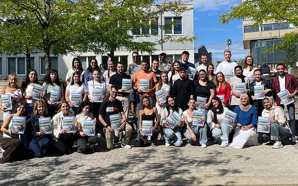
    • specialized diagnostics: specialist for complex diagnostic reporting at pathology institutes and in private laboratories

    • management roles in laboratories

      • self-employment: working as a freelance biomedical scientist with a specialized portfolio

      • science: participation in clinical trials and research projects in cellular diagnostics


        News

        all news
        Eine große Gruppe junger Menschen posiert auf Stiegen, sie halten bunte Mappen in der Hand. Im Hintergrund ist ein Gebäude sowie Himmel zu sehen.
        17. September 2025

        Seven countries, one event

        The Erasmus+ Blended Intensive Programme of the Radiological Technology bachelor's program brought international students and lecturers together in Vienna for the third time.

        • Health Sciences
        • Networking
        • Mobility

        17. October 2024

        3D printing, mixed reality and AI at ViMiTech 2024

        • Health Sciences
        SHOW MORE

        Vernetzen mit Absolvent*innen und Organisationen

        We work closely with the Vienna Healthcare Group, private laboratories, the Medical University of Vienna, European universities, the professional association biomed austria, and healthcare institutions. This provides you with opportunities for practical learning phases, job searches, or participation in research and development activities. You will complete the extensive practical learning phases at locations including clinics within the Vienna Healthcare Group. Many of our partnerships are listed on the Campusnetzwerk platform. It’s always worth taking a look—it might lead you to a new job or an interesting event hosted by our Cooperation Partners!

        Research projects and activities


        Downloads

        Infofolder Health Sciences (content in German)
        pdf, 1,024 KB

        Contact

        Head of program

        Administration

        Andreas Moser

        +43-1-6066877-4130

        Susanne Flögel
        +43-1-6066877-1782

        Favoritenstraße 226, D.1.33 (building plan)
        1100 Wien

        cytology@hcw.ac.at

        Office hours
        Thursday 2.00 pm to 5.00 pm
        Friday 9.00 am to 3.00 pm
        or by appointment

         

        You want to know more? We will help you.


        Have you found all the information you are looking for?

        • Yes
        • No